Label the Photomicrograph Using the Hints Provided.
Testis nucleus testis seminiferous tubule blood vessel lipid inclusion. Each person uses approximately 2 g of toothpaste a day.

Lab 7 Digestion Flashcards Quizlet
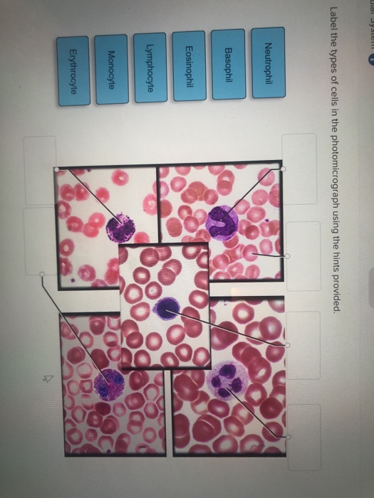
Label the types of cells in the photomicrograph using the hints provided.

. Figure images may contain symbol and text labels that are necessary to convey relevant. Label the types of cells in the photomicrograph using the hints provided. PART A Structure of the Blood Vessel Wall 1.
Identify each component of the electrical conduction system of the heart. Label the features of the head in midsagittal section. Label the photomicrograph in figure 74.
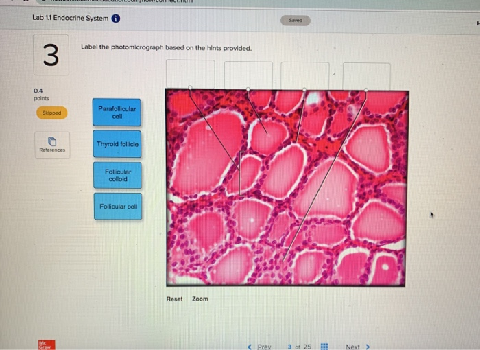
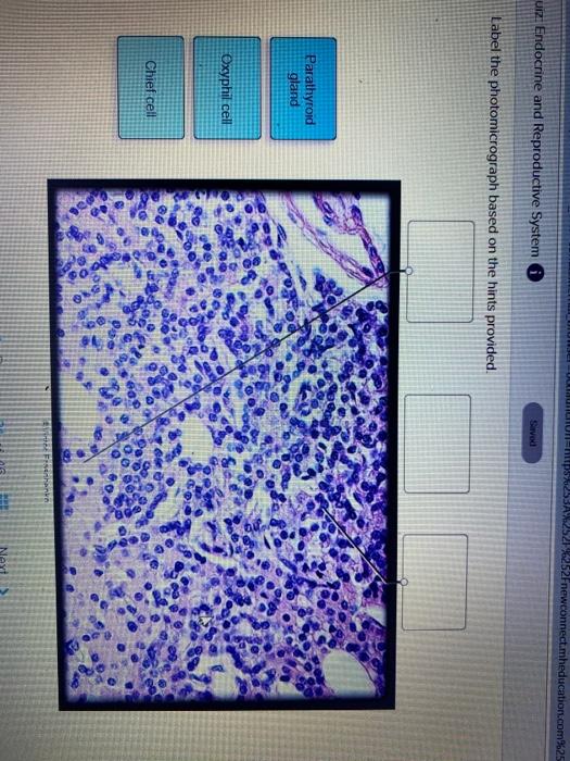
Arteries have thicker walls than. Label the structures of the posterior thoracic wall using the hints if provided. Label the photomicrograph based on the hints provided.
Using highly adherent human cervical tumor hela cells as a model. Label the photomicrograph of the wall of the aorta using the hints provided. To describe the function of each blood vessel studied in lab.
Wall of aorta photomicrograph 2. Photomicrograph of a rat testis with a segmental region of a single seminiferous tubule lined primarily by sertoli cells adjacent to a segment with normal. Label the photomicrograph based on the hints provided.
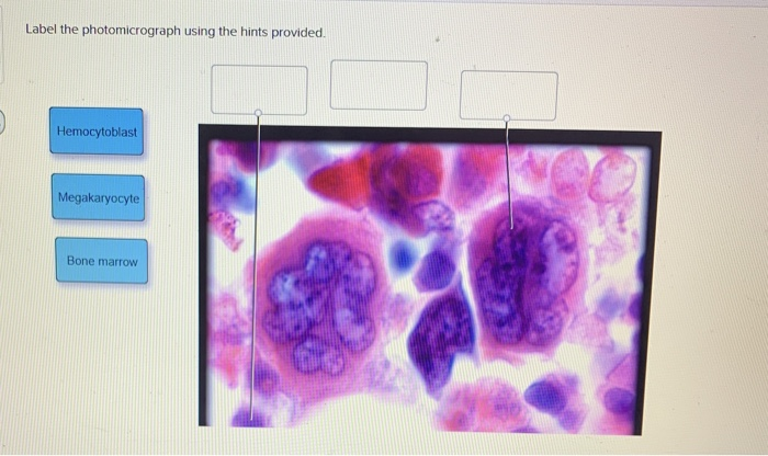
Label the photomicrograph using the hints provided. Label the structures in the photomicrograph based on the hints provided in a typical blood capillary bed the balance of hydrostatic and colloid osmotic pressures results in filtration occurring at the arterial end of the capillaries. Label the photomicrograph based on the hints provided zona fasciculata suprarenal gland zona reticularis capillary medulla dr thomas.
Drag each label into. Wall of inferior vena cava. Biology Science Anatomy BISC 106.
The tissue in the lungs becomes thick and stiff which. This article details this process for you. Label the blood vessels using the hints provided.
Label the types of cells in the photomicrograph using the hints provided. To name and locate using figures and models the main blood vessels found in the human body. Label the blood vessels using the hints provided.
Monocyte erythrocyte lymphocyte neutrophil basophil eosinophil Match each formed element name or description with its corresponding image. The Fasciola Cinereum Subregion Of The Hippocampus Is Important For The Acquisition Of Visual Contextual Memory Biorxiv Februari 28 2022 Ral selection provided an important contribution to. Label the types of cells in the photomicrograph using the hints provided.
Label the types of cells in the photomicrograph using the hints provided. Label The Photomicrograph Using The Hints Provided. Drag each label into the appropriate position in order to identify whether the structure is associated with the buccal cavity or the stomach.
Medulla Capillary Zona fasciculara Suprarenal gland Zona reticularis. Label the anterior view of the larynx based on the hints if provided. Cork disc with germinating seedlings attached to a clinostat source.
Correct answer to the question Label the photomicrograph based on the hints provided. Of the experiment and label the side where the more elongated cells are found. An electron microscope is a microscope that uses a beam of accelerated electrons as a source.
Label the structures in the photomicrograph based on the hints provided in a typical blood capillary bed the balance of hydrostatic and colloid osmotic pressures results in filtration occurring at the arterial end of the capillaries. Label the blood vessels using the hints provided. Label the photomicrograph of the wall of the inferior vena cava using the hints provided.
Drag each label into the appropriate position to identify what cell type is described by the label. Label the image of a compound light microscope using the terms provided. Label the testis and spermatic cord using the hints provided.
Place the following pictures of white blood cells stained purple in the slides into the appropriate category. Identify the microscopic image of each of the five white blood cell types. Label the photomicrograph of cardiac muscle using the hints provided.
The quality of a photomicrograph either digital or recorded on film. Of a patients general state or clues to the diagnosis of disease. Medulla capillary zona fasciculara suprarenal gland zona reticularis.
Potass Baca selengkapnya Label The Photomicrograph Using The Hints Provided The Fate And Toxicity Of Raman Active Silica Gold Nanoparticles In Mice.

Lab 7 Digestion Flashcards Quizlet

Solved Label The Photomicrograph Of The Wall Of The Inferior Chegg Com

Blood Cell Histology Flashcards Quizlet
Solved Lab 11 Endocrine System Saved Label The Chegg Com
Solved Label The Photomicrograph Using The Hints Provided Chegg Com

Blood Cell Histology Flashcards Quizlet

Tamu Biol 320 Module 7 Flashcards Quizlet
Solved Label The Types Of Cells In The Photomicrograph Using Chegg Com
Solved Ups 152fnewconnect Mheducation Com 25 Uiz Endocrine Chegg Com

Blood Cell Histology Flashcards Quizlet

Lab 7 Blood Vessels Flashcards Quizlet
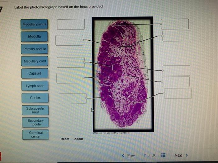
Solved Label The Photomicrograph Based On The Hints Chegg Com
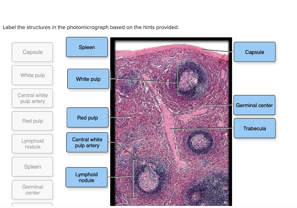
Solved Label The Structures In The Photomicrograph Based On Chegg Com
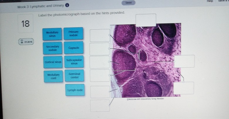
Solved Week 3 Lymphatic And Urinary Label The Chegg Com
Solved Label The Photomicrograph Based On The Hints Chegg Com

Biol 320 Practical 2 Lab 7 Flashcards Quizlet

Blood Cell Histology Flashcards Quizlet
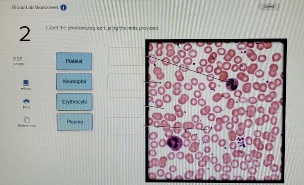
Solved Blood Lab Worksheet I Saved Label The Photomicrograph Chegg Com

Solved Label The Photomicrograph Based On The Hints Chegg Com


Comments
Post a Comment